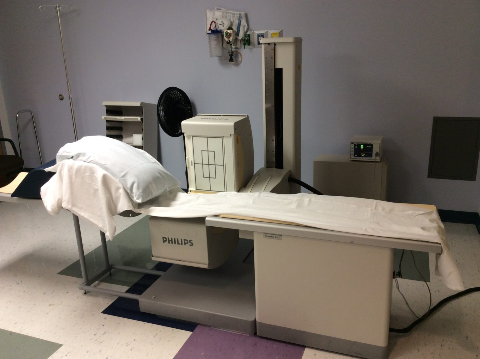
Nuclear Scan
A nuclear scan is a type of imaging test that shows how well an organ is functioning.

During a nuclear scan, a small amount of radioactive material (called a tracer or isotope) is injected into the bloodstream. The organ being studied absorbs the tracer, and a special camera detects the radiation it gives off. By moving around the body, the camera captures images from different angles, and a computer combines these into detailed pictures of the organ and its circulation.
Why the Test is Performed
Nuclear scans are used to evaluate how well organs such as the heart, lungs, thyroid, bones, liver, or kidneys are working. Unlike many other imaging tests that focus only on structure, nuclear scans show both function and blood flow. They help providers diagnose conditions like cancer, infections, fractures, heart disease, or thyroid disorders, and they can also track how well treatments are working.
It's important to note that nuclear scans are not suitable for all patients. They may not be advised for pregnant or breastfeeding women, or in people with certain medical conditions. Consult with your healthcare provider to determine if a nuclear scan is appropriate for you.
What to Expect
Before the scan, a technologist will inject the radioactive tracer into your arm. You may be asked to wait a short time while your body absorbs the material. During the scan, you’ll lie on a table while the camera moves slowly around you. The procedure is painless and typically lasts between 30 minutes and 2 hours, depending on the type of scan. Afterward, you can return to normal activities. The tracer leaves your body naturally, usually within a day or two, and drinking fluids can help speed up this process.
