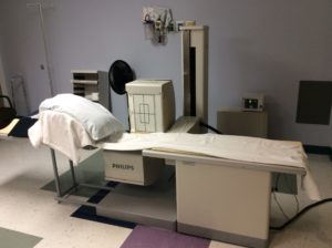
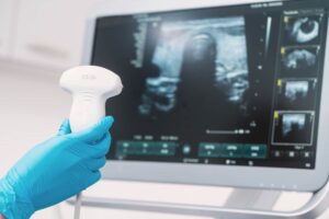
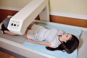
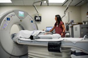
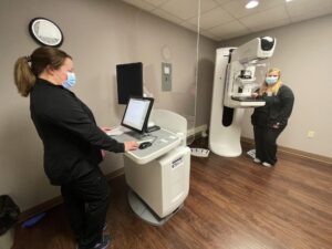
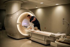
Radiology & Imaging

October 30, 2017

Radiology & Imaging

Newman Regional Health’s Radiology Department is fully equipped with state-of-the-art imaging technology. Our advanced medical imaging services assist our providers in diagnosing medical problems quickly and formulating effective treatment plans. All images are interpreted by a board-certified radiologist, who works closely with the referring physician and nursing team.

CT Scan

Computed tomography (CT), also known as a “CAT scan,” is a medical diagnostic tool that creates images of internal structures within the human body. CT scans aid physicians in diagnosing disease, viewing internal abnormalities, and assessing the extent of trauma. Newman Regional Health is equipped with a State of the Art Toshiba 160 Slice CT Scanner that provides highly detailed images of the human body.

The Toshiba 160 slice CT scanner non-invasively assists physicians in the diagnosis of a variety of anatomical areas, including spine, head, abdomen and chest. The Toshiba 160 slice CT scanner allows providers to simultaneously capture multiple wafer-thin images of a patient’s anatomy within seconds, shaving precious minutes off the exam time.

The Toshiba 160 Slice CT system compliments and extends the high range of quality diagnostic services that Newman Regional Health offers the residents of Emporia and the surrounding area. The Toshiba 160 Slice CT Scanner provides our providers with vastly improved image quality, and entry into new applications such as CT angiography and an overall enhanced diagnostic confidence on all studies.

CT Temporal Bones
CT IAC (Internal Auditory Canal)
CT Mandible
CT Facial Bones
CT Orbits
CT Sinuses
CT Orthopedic
CT Head
CT Low-dose scan for lung cancer screening
CT Neck Soft Tissue
CT Extremities
CT Spine
CT Urogram
CT Chest
CT Urogram
CT Stone Search
CT Abdomen
CT Pelvis
CT Angiogram)

MRI (Magnetic Resonance Imaging)

Magnetic resonance imaging (MRI) produces detailed images of the body’s organs, tissues and joints without patient discomfort or exposure to radiation. MRI uses a powerful magnetic field and radiofrequency to produce computerized images. MRI is especially useful for imaging the brain, spine, and joints.

For providers, MRI is advantageous in determining soft tissue disorders, including cartilage and ligament tears and herniated disks, but it also can be helpful in diagnosing bone disorders such as tumors.

Designed with the patient in mind, Newman Regional Health’s Philips Ingenia  1.5 Tesla ultra-short bore MRI system is the shortest, most open high field scanner available. The state-of-the-art digital clarity and speed of this machine helps physicians diagnose with confidence and work productively. Newman’s MRI is comfortable for patients because of its wide bore design. The increased width of the machine opening decreases claustrophobic feelings and the constant ability to communicate with the MRI technologist helps ease patient’s fears during their exam. These two features combined with the decreased scan times of the Ingenia system allow for a more tolerable environment for all patients, not just those with claustrophobia.

Newman Regional Health’s MRI department is able to do a wide variety of exams to help physicians with clinical diagnosis.  The Ingenia is a well-rounded machine that has the capability to make exceptional images of all body parts.  Your doctor may order the MRI with an IV dye injection, this can also be easily accomplished at our facility.  

Functional MRI
Cardiac MRI
MRI of the Breast
Magnetic Resonance Venography (MRV)
Non-Contrast MRA

Nuclear Scans

A nuclear scan shows how well an organ functions. The procedure begins with the injection of small amounts of radioactive trace material (isotopes). The organ absorbs the radioactive material. The special camera on the nuclear scan detects the radiation in the organ. The camera moves at different angles and sends signals to a computer that constructs an image of an organ and its circulation. Nuclear scans help providers diagnose many conditions, including cancer.

Ultrasound

Ultrasound is a non-invasive procedure that uses high frequency sound waves to create images. During ultrasound, a technician moves a small device on the skin. The device transmits signals that a computer converts into moving images. The image is displayed on a computer screen.

There are many uses for ultrasound including viewing a developing fetus, abdominal organ, thyroid, prostate, heart function, and blood flow through major vessels.

PET Scans

Doctors use PET scans to get more data about how body organs are functioning. PET scans may be performed together with a computerized axial tomography (CAT) scan that provides an image of the organ. PET scans provide a clear view of how the organs are working at the cellular level and if they have been damaged. The scan helps doctors determine effective treatment options.

PET scans are commonly used to diagnose heart conditions, help doctors determine appropriate cancer treatment, help in diagnosing Alzheimer’s disease and brain disorders.

They also can provide data for medical research.

PET/CT Scans are performed in a special MOBILE FACILITY that travels around Kansas on a specific schedule. Appointments are made through your care provider and generally are performed on Fridays.

Meet the Providers

Andy Legako, DO
Andy Legako, DO
Frank Yackovich, MD
Frank Yackovich, MD

Connect With Us

Newman Regional Health
1201 W 12th Avenue
Emporia, KS 66801
620-343-6800
Newman Medical Plaza
1301 W 12th Avenue
Emporia, KS 66801